Newsletters

Oxygen Imaging of Cells for Viability Assessment

Cell viability is an essential measurement for cell therapy, tissue-engineered medical products (TEMPs), drug development, and many other biological processes and products. These systems rely on viable, healthy, and functional cells to work as intended.

However, current assay-based methods for assessing cells are destructive and inadequate. In our recently published paper in npj imaging, we report partial oxygen pressure (pO2) maps of cells in an environmentally controlled multi-well plate as a new method to obtain a 3D, longitudinal assessment of cells without destroying them in the process. The data presented here could be useful to every biological lab. 

Using oxygen imaging of adherent and non-adherent cells seeded in an environment-controlled multi-well plate incubator-resonator, we demonstrated that:

  • We can assess cells’ metabolic activities longitudinally and in 3D without destroying them in the process for up to 24 hours.
  • We can distinguish between live and dead cells.
  • The cells can be hypoxic even in the incubator.
  • We can assess cells’ metabolic activity in a culture medium, but more importantly, when also seeded in a hydrogel.
  • The oxygen concentration of wells with low cell density is high and decreases exponentially with increasing cell seeding density. However, it changes with time as cells become dormant, differentiate, propagate, or die. Therefore, cell density is an important factor to control when using them for cell therapy and tissue engineering.
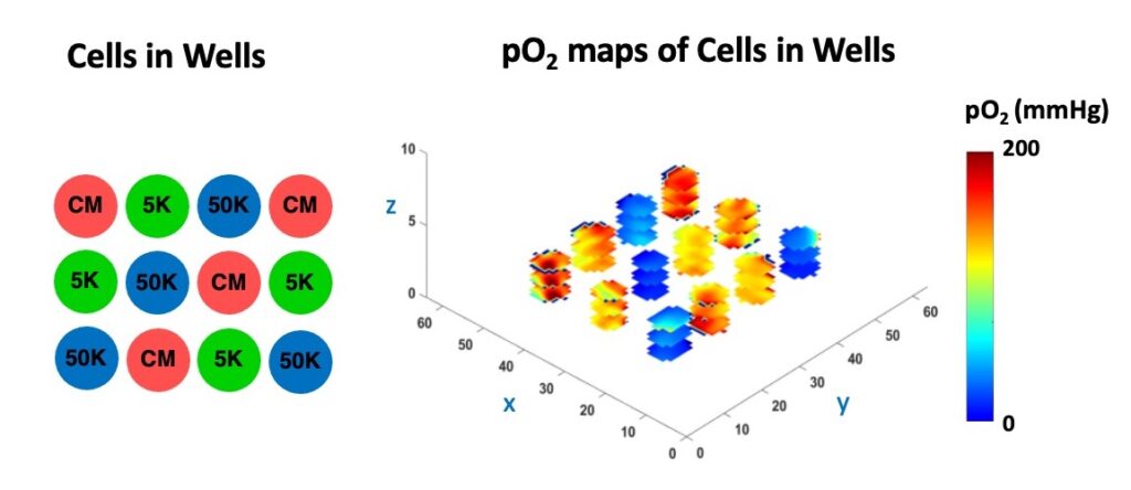
The figure below shows the pOmaps and box plot of live HEK 293 cells seeded with different densities (low oxygen means the cells are alive and actively consuming oxygen, while dead cells do not).

The following figure shows how the average pO2 evolved over the course of 4 hours: the mean pO2 of 50K cells matched the pO2 of wells with 100K cells, where cell dormancy or cell death was probably happening because of overcrowding. The pO2 for 25 K cells dropped over time, indicating increased metabolic activity in these wells.

An important challenge in evaluating tissue-engineered medical products arises when cells are implanted into a hydrogel scaffold because there is no method available to monitor their metabolic activity without disrupting the structure. In the figure below, we show that the quantitative estimate of cell loss for cells seeded in a biomaterial can be obtained using oxygen imaging.

In the Figure below, we demonstrate the 3D cell viability assessment using oxygen imaging.

This is the first study showing nondestructive, 3D, longitudinal cell viability assessment using 3D oxygen imaging and it may impact cell therapy, tissue engineering, regenerative medicine, and other fields. All experiments were performed using O2M's preclinical oxygen imager, JIVA-25®.

This is an important assay for every biological lab, as the cells subjected to different oxygen concentrations can lead to vastly different outcomes!

Click here to access the full paper and learn more!

Oxygen Imaging of Cells for Viability Assessment Read More »

Quantification of Blood-Brain Barrier Leakage and Brain Oxygen Through EPR Imaging

Did you know that BBB leakage can be quantified non-invasively with EPR Imaging?

In a healthy brain, the EPR imaging contrast agent, trityl OXO71, is blocked by the blood-brain barrier (BBB), but guess what, when the BBB is compromised because of any neurological issue, trityl can enter and light up the brain. Therefore, EPR imaging provides a straightforward way to image and quantify BBB when it's compromised. Moreover, when BBB is damaged, we can map three-dimensionally the brain partial oxygen pressure (pO2), which has not been explored before because of lack of technologies.

 The corresponding publication titled "Assessment of blood-brain barrier leakage and brain oxygenation in Connexin-32 knockout mice with systemic neuroinflammation using pulse electron paramagnetic resonance imaging techniques" was published in 2024 in Magnetic Resonance in Medicine (MRM) journal - (Volume 91, pages 2519-2531).

Click here to learn more or contact us is you are interested in a collaboration!

fig_brain

Quantification of Blood-Brain Barrier Leakage and Brain Oxygen Through EPR Imaging Read More »

Accelerated EPR Imaging using Deep Learning Denoising

As OXO71-based Electron Paramagnetic Resonance Oxygen Imaging (EPROI) transitions from small to large animal models, and ultimately to humans, various challenges must be addressed. Long acquisition times and low SNR of individual scan pose significant challenges.

In the paper "Accelerated EPR Imaging using Deep Learning Denoising" , published in Magnetic Resonance in Medicine journal, in collaboration with Dr. McMillan from University of Wisconsin-Madison, we tried to tackle some of these issues using deep learning techniques. Our approach leverages a UNet enhanced with residual units, followed by two trainable joint bilateral filters (UNet+JBF2)The results are extremely encouraging:

  • 10× reduction in number of averages (from 150 to just 15 shots) while maintaining SNR
  • Noise levels comparable between the filtered 15-shot acquisition and the original 150-shot acquisition (see Figures d, e, below).
  • 3x improvement in image quality for 150-shots acquisitions, boosting the SNR from 240 to 666 (p-value = 0.0074).
fig2

Even more excitingly, our model generalizes to oxygen maps. It successfully denoises amplitude maps without distorting pO2 estimations (Figure a, below) and enhances SNR by 2.4 fold for in vivo tumor pO2 maps (Figure b, below).

Click here or on the images to learn more!

fig3

Accelerated EPR Imaging using Deep Learning Denoising Read More »

Mouse Abdominal pO2 Imaging and Assessment of Subcutaneously Implanted Cell Therapy Devices

Type 1 diabetes (T1D) is an autoimmune disease that leads to the loss of insulin-producing pancreatic beta cells. Beta cell replacement devices or bioartificial pancreas (BAP) have shown promise in curing T1D and providing long-term insulin independence without the need for immunosuppressants. However, hypoxia in BAP devices can damage the cells and limit device dimensions. Noninvasive in vivo oxygen imaging assessment of implanted BAP devices will provide the necessary early feedback and improve the chances of success. In the study titled In Vivo Mouse Abdominal Oxygen Imaging and Assessment of Subcutaneously Implanted Beta Cell Replacement Devices, published in Molecular Imaging and Biology journal, we optimized mouse abdominal oxygen imaging using our core technique, electron paramagnetic resonance oxygen imaging (EPROI) and our novel oxygen imager, JIVA-25®, to assess BAP devices in vivoThis study is part II of our previously published study demonstrating in vitro oxygen assessment of BAP devices. fig1

We demonstrated proof-of-concept pO2 imaging of two subcutaneously implanted BAP devices: an agarose-based cylindrical device (OxySite) implanted in streptozotocin (STZ)-induced diabetic animals and a commercially available cell encapsulation device (TheraCyte) implanted in non-diabetic animals. Check out the full study here.

fig2

Mouse Abdominal pO2 Imaging and Assessment of Subcutaneously Implanted Cell Therapy Devices Read More »

Oxygen Imaging and Tumor Radiotherapy

Cancer is a major public health problem worldwide and is the second leading cause of death in the United States. About two-thirds of cancer patients are treated with radiation, either alone or in combination with immunotherapy, chemotherapy, or surgery. It is known that hypoxia or low partial oxygen pressure (pO2) causes resistance to radiation therapy, immunotherapy, and chemotherapy.

Solid tumors treated with radiotherapy often experience decreased oxygen delivery.  This early tumor response to radiotherapy can be monitored by measuring the pO2 in the tumor microenvironment.  These in vivo pO2 measurements can be accomplished using the JIVA-25™ Electron Paramagnetic Resonance Oxygen Imaging (EPROI) instrument from O2M Technologies.  During EPROI studies, a gas anesthetic is often used to immobilize the mouse.  Unfortunately, a gas anesthetic that uses medical grade air (21% O2) or 100% O2 breathing gas can alter the measured pO2 in the tumor, potentially complicating the interpretation of the imaging results.

Figure 1.  Oxygen Enhanced EPROI.  The workflow acquires anatomical MR images, two pOmaps with 21% O2 breathing gas, and two pO2 maps of 100% O2 breathing gas.  The test-retest shows outstanding precision of 3.1 Torr with both breathing gases.

Prof. Marty Pagel and graduate student Tianzhe Li at the MD Anderson Cancer Center have exploited this potential pitfall to develop a new biomarker for assessing early tumor response to radiotherapy.  They have measured ΔpO2, which is the difference in pO2 measured with 21% O2 and 100% O2 breathing gases.  As an analogy, a car with a ¼-full gas tank has an available capacity of ¾-empty tank.  ΔpO2 represents the available capacity of a tumor to take more O2.  To demonstrate the value of this new biomarker, Li and Pagel have developed an Oxygen Enhanced (OE)-EPROI protocol that interperitoneally administers OX071 agent, acquires MR images for anatomical reference, and then obtains pO­2 maps with 21% O2 and 100% O2 breathing gases (Figure 1).  They have applied their protocol to study the early response of a Colo357 model treated with 10 Gy radiotherapy (Figure 2).

Figure 2.  Oxygen Enhanced (OE)-EPROI showing a significant decrease in ΔpO2.

Their results have shown that radiotherapy caused a large and significant decrease in ΔpO2 (Table 1). For comparison, radiotherapy caused a small, statistically insignificant decrease in tumor pO2 with 21% O2 breathing gas.  Similarly, radiotherapy caused a larger yet still statistically insignificant decrease in tumor pO2 with 100% O2 breathing gas.  Furthermore, ΔpO2 showed the greatest effect size, known as the change in a biomarker relative to its variance, demonstrating that ΔpO2 was most effective in evaluating an early tumor response to radiotherapy.  This OE-EPROI protocol can provide a new diagnostic method for evaluating early response to radiotherapy.

Table 1. OE-EPROI of the Colo357 model undergoing radiotherapy (n=9)

  1. differences determined for each mouse, and not based on the averages of the groups.
  2. p < 0.01 is considered to be statistically significant, from a two-tailed Student’s T-Test assuming equal variances
  3. Effect size: |{(average post-pre) / (average standard deviation of post and pre)}|

Prof. Marty Pagel leads the Contrast Agent Molecular Engineering Laboratory (CAMEL) at the MD Anderson Cancer Center, Houston TX, USA.  CAMEL evaluates hypoxia, acidosis, enzyme activity and vascular perfusion in the tumor microenvironment, using a variety of molecular imaging methods.  More specifically, CAMEL uses EPROI to evaluate chemotherapy, immunotherapy, radiotherapy and radiosensitizers in small animal models of cancer and models of wound healing.

Learn more about EPR Oxygen Imaging

Oxygen Imaging and Tumor Radiotherapy Read More »

Oxygen Sensitive Trityl Radical OX071

Magnetic resonance imaging (MRI) and electron paramagnetic resonance oxygen imaging (eMRI or EPROI) are fundamentally based upon the same principles, however a key difference is the resonant species. MRI aligns water protons, that are abundant in the body, in a magnetic field to produce exquisite anatomical images whereas eMRI relies on aligning electron spins. Unlike the ubiquity of protons, unpaired electrons sensitive to oxygen are rarely found in living organisms. A nontoxic contrast agents, with high specificity and sensitivity to detect molecular oxygen, is a necessity for EPR-based oxygen imaging; and now, it is being synthesized by O2M Technologies. Introducing incredible molecule: OX071!

Tetrathiatriarylmethyl or “trityl” radicals are not unfamiliar in MRI applications. They have been used as a polarizing agent. Previous versions of trityls, such as the largely used Finland trityl (FT), were unsuitable for in vivo experimentation. Its lipophilic core has a propensity to aggregate with plasma proteins, resulting in some amount of toxicity. In the next iteration, trityl OX063, 12 methyl groups in FT are replaced with hydroxyethyl groups, which greatly increased it’s hydrophilicity. The result is a completely non-toxic, injectable probe. OX063 is used a polarizing agent in 13C MRI.

The latest evolution of trityl is OX071, a partially deuterated analogue of OX063. Deuterium is EPR-silent, which eliminates a source of hyperfine coupling with trityl’s radical center. OX071 retains all the benefits of it’s predecessor while also showing a higher oxygen sensitivity, a higher signal-to-noise ratio, and under physiological conditions, a single-line EPR spectrum with ultra-narrow linewidth. OX071 is especially sensitive in low oxygen conditions. Even a small amount of OX071 will generate signal. The probe can be delivered intravenously, intraperitonially or intratumorally. OX071 is cleared from the animal in 10-30 minutes virtually unchanged.

All these properties make OX071 the probe of choice for a wide array of in vivo EPR oximetry experiments. OX071 has already been published in applications of solid tumor oximetry, viability studies in highly oxygen dependent cell populations like transplanted islets of Langerhansbioscaffold device development, and much more. Reach out to O2M for OX063 or OX071 in your experiments!

Oxygen Sensitive Trityl Radical OX071 Read More »

Finding the Best Site for Tissue Graft Implantation

Oxygen sensitive LiPc probes implanted within the right dorsum subcutaneously, reporting oxygen tension  over 6 weeks. Data obtained by JIVA-25™ oxygen imager. Oxygen sensitive LiPc probes implanted within the right dorsum subcutaneously, reporting oxygen tension over 6 weeks. Data obtained by JIVA-25™ oxygen imager.

Tissue engineered grafts and cell encapsulation devices have significant potential to improve human health. The site of transplantation is a factor that has profound effect on graft survival. The ideal chosen site should be easy to access for implantation and retrieval, while providing adequate diffusion of nutrients and oxygen for support of cell survival until vascularization is established. The two most commonly used sites for implantation of tissue grafts and cell encapsulation devices are subcutaneous (SC) and intraperitoneal (IP). Evaluating the native oxygenation in these sites is valuable information towards understanding the physiological milieu of implants in vivo.

O2M’s recent peer-reviewed publication in Tissue Engineering Part C: Methods presents the first ever solid probe oxygen imaging, reporting pO2 values, of subcutaneous and intraperitoneal spaces in mice. We observe that solid probe oxygen imaging is a technology that offers researchers oxygenation data in vivo without needing further invasive measures or sacrifice. This study was funded by Juvenile Diabetes Research Foundation (JDRF) and will help scientists develop better therapies for type I diabetes as well as other health conditions.

Visit O2M’s Publication Page to learn about latest research in oxygen imaging.

O2M is at BioFAB and TERMIS!

Come see us in Manchester, New Hampshire!

Spring 2022 Meeting in the Millyard
June 7 – June 9 O2M will be presenting our latest research at the Poster Session
Wednesday June 8, Currier Museum, 5:30pm – 8:00pm

“In Vitro and In Vivo Oxygen Imaging of Islet Encapsulation Devices: Lessons Learned and Path Forward”

Come See Us in Toronto!

TERMIS-AM 2022
July 10 – July 13
Booth #107

Are you at TERMIS 2022? Make sure your plans include a visit to our booth in the Exhibition Hall. We hope to see you there!

O2M is Presenting at TERMIS! Keynote Talk by Dr. Mrignayani Kotecha
“In Vitro and In Vivo Oxygen Imaging assessment of Islet Transplantation Devices”
Sessions 7, Non-invasive Imaging and Analysis of Engineered Tissues
Wednesday, July 13, 8:00 AM – 8:30 AM Poster Presentation by Team O2M
“In Vivo Oxygen Imaging Of Oxygen Generating Cellular Implant Devices”
Poster Session 3
Tuesday, July 12, 4:30pm – 6:00pm

Upcoming O2M Webinar

1e1bb326-f53f-0b03-88d8-a0bc20ae9257 Register for Webinar

Moderator: Dr. Martyna Elas, Jagiellonian University, Krakow, Poland

About the Speaker:
Dr. Mark “Marty” Pagel has focused on molecular imaging research during the last 20 years in industry and academia.  He is a Professor in the Departments of Cancer Systems Imaging and Imaging Physics at the University of Texas MD Anderson Cancer Center.  In addition, Dr. Pagel has held leadership positions in professional societies, funding agencies, and scientific journals that focus on molecular imaging.  Dr. Pagel’s current research focuses on CEST MRI, PET/MRI, photoacoustic imaging, and EPR imaging, for studies of tumor hypoxia, acidosis, vascular perfusion and enzyme activity, with mouse models of cancer and for clinical trials with cancer patients.

Abstract: Previous studies with EPR imaging have shown that measurements of tumor pO2 can indicate the status of hypoxia in the tumor microenvironment, which can be used to predict response to radiation therapy.  To build on these previous studies, we are developing Oxygen Enhanced (OE) EPR imaging that challenges a pre-clinical tumor model with 21% O2 (medical grade air) and 100% O2 in the anesthetic carrier gas, which can be a useful biomarker for evaluating early response to radiation therapy.  In addition, we are developing Dynamic Contrast Enhanced (DCE) EPR imaging that measures tumor vascular perfusion, as a complimentary biomarker for evaluating early response to cancer treatment.  This presentation will also discuss our ongoing research studies that show how tumor hypoxia causes resistance to immunotherapy, and how reducing hypoxia can improve tumor control with immune checkpoint blockade.

Finding the Best Site for Tissue Graft Implantation Read More »

Biomaterial Assessment Using Oxygen Imaging

Biomaterials that host live cells must overcome the crucial hurdle of sustaining sufficient oxygenation for cell survival. Several approaches for improving oxygenation within the biomaterials have been developed and investigated for improved cell survival in artificial tissue constructs. Regardless of the method of addressing the oxygen needs of the cells, all three-dimensional constructs require in situ pO2 assessment in vitro and in vivo to gauge the success of the approach. In addition, in vivo models can have very divergent results because of inherent variability between the animals, which demands a greater understanding for the breadth of variation in normal and diseased states.  O2M’s oxygen imaging technology can improve the outcome of cell and tissue engineering by providing real time in vitro and in vivo oxygen maps for improved therapy outcome.
O2M’s “Oxygen Measurement Core” partners with research labs to provide critical oxygenation and associated biological data to accelerate their development of better biomaterials, cell replacement devices, and tissue constructs. Recent publications from these partnerships have been published at Nature CommunicationsScience Advances, and Journal of Biomedical Materials Research.
Reach out to O2M to find out how oxygen imaging can advance the development of your biomaterial research!

O2M is at SFB!

Come see us in Baltimore! If you are at SFB 2022, make sure your plans include a visit to us. Get a hands-on demonstration with our in-vivo oxygen imager, JIVA-25™. We hope to see you there!

O2M is Presenting at SFB!
Come learn about our latest research!

Biomaterial-Tissue Interaction
Thursday, April 28, 11:00am – 11:15am
In Vivo pO2 Assessment of Implantation Site: SubQ vs IP
Oral Presentation by CEO Dr. Mrignayani Kotecha

Biomaterials for Pancreatic Islet Replacement and Immune Tolerance in T1D
Saturday, April 30, 12:15-12:30pm
In Vivo pO2 Measurement of Islet Encapsulation Devices in Oxygen Measurement Core
Oral Presentation by CEO Dr. Mrignayani Kotecha

Biomaterial Assessment Using Oxygen Imaging Read More »

Accelerating Towards a Cure for Type 1 Diabetes

Type 1 diabetes mellitus (T1D) is an autoimmune disease causing insufficient or no production of insulin from the pancreas; this impedes glucose metabolism, generating several life-long health sequelae in nearly every organ system. Tools such as automated exogenous insulin delivery systems and constant glucometers, alongside diligent self-care, do improve outcomes. However, additional therapies are needed to improve the management of therapeutic goals and eliminate the risks or consequences of wide swings in glycemia. Transplantation of human islets  has offered a proof-of-concept for beta-cell replacement therapy. But a number of hurdles remain in realizing the full potential of beta-cell replacement therapy, one of which being ensuring an adequate supply of nutrients and oxygen to the islet graft.

photo_2022-01-06_16-25-28

In response, Juvenile Diabetes Research Foundation (JDRF) is collaborating with a wide spectrum of partners to develop a replacement product using an unlimited source of beta cells without needing broad spectrum immunosuppression. A critical challenge is to protect beta cells from their host’s immune system, while still preserving consistent gas permeability, and permitting glucose-responsive insulin secretion. Novel designs are emerging that try to strike this delicate balance between diffusion, perfusion, and protection (see figure). Non-invasive tools that can directly map oxygen levels in vivo could prove very useful in evaluating and optimizing new technologies that extend the viability of beta-cell implants. In an effort to accelerate the development of beta cell replacement therapies, JDRF is supporting O2M Technologies’ “Oxygen Measurement Core” to test the therapeutic potential of implantable cell delivery systems. The technology developed by O2M has already proven to be successful in providing oxygen maps of islet encapsulation devices in vitro and in mice.

JDRF is committed to achieving their vision of “a world without T1D.” By connecting multidisciplinary collaborations and investing in diverse strategies, JDRF is closing research gaps. See a discussion by VP of Research Dr. Esther Latres on how JDRF is accelerating towards a cure for T1D.

Accelerating Towards a Cure for Type 1 Diabetes Read More »

Scroll to Top