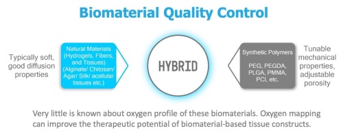
Biomaterials that host live cells must overcome the crucial hurdle of sustaining sufficient oxygenation for cell survival. Several approaches for improving oxygenation within the biomaterials have been developed and investigated for improved cell survival in artificial tissue constructs. Regardless of the method of addressing the oxygen needs of the cells, all three-dimensional constructs require in situ pO2 assessment in vitro and in vivo to gauge the success of the approach. In addition, in vivo models can have very divergent results because of inherent variability between the animals, which demands a greater understanding for the breadth of variation in normal and diseased states. O2M’s oxygen imaging technology can improve the outcome of cell and tissue engineering by providing real time in vitro and in vivo oxygen maps for improved therapy outcome.


O2M’s “Oxygen Measurement Core” partners with research labs to provide critical oxygenation and associated biological data to accelerate their development of better biomaterials, cell replacement devices, and tissue constructs. Recent publications from these partnerships have been published at Nature Communications, Science Advances, and Journal of Biomedical Materials Research.
Reach out to O2M to find out how oxygen imaging can advance the development of your biomaterial research!
O2M is at SFB!

Come see us in Baltimore! If you are at SFB 2022, make sure your plans include a visit to us. Get a hands-on demonstration with our in-vivo oxygen imager, JIVA-25™. We hope to see you there!
O2M is Presenting at SFB!
Come learn about our latest research!
Biomaterial-Tissue Interaction
Thursday, April 28, 11:00am – 11:15am
“In Vivo pO2 Assessment of Implantation Site: SubQ vs IP”
Oral Presentation by CEO Dr. Mrignayani Kotecha
Biomaterials for Pancreatic Islet Replacement and Immune Tolerance in T1D
Saturday, April 30, 12:15-12:30pm
“In Vivo pO2 Measurement of Islet Encapsulation Devices in Oxygen Measurement Core”
Oral Presentation by CEO Dr. Mrignayani Kotecha
|
|
