This spring, O2M is hosting a webinar mini-series on cancer hypoxia! Join us next month for a conversation with Duke University team Drs. Greg Palmer, Yvone Mowery, & Ashlyn Rickard, investigating how oxygen imaging can optimize the dose and timing of an FDA approved drug papaverine in hypoxia reduction and improved radiation treatment. Then in June hear Dr. Marty Pagel from the University of Texas MD Anderson Cancer Center, exploring tumor microenvironment using oxygen imaging.
Reach out to O2M Technologies to discuss how oxygen imaging can support your cancer research!
May 2022 O2M Webinars

Moderator: Prof. Marty Pagel, MD Anderson Cancer Center
About the Speakers:
Greg Palmer is currently an Associate Professor in the Department of Radiation Oncology, Cancer Biology Division at Duke University Medical Center. His primary research focus has been identifying and exploiting the changes in absorption, scattering, and fluorescence properties of tissue associated with cancer progression and therapeutic response.
Yvonne M. Mowery, MD, PhD, DABR is a physician scientist and the Butler Harris Assistant Professor or Radiation Oncology at Duke University, specializing in treating head and neck cancer with radiotherapy and studying novel combinations of radiation with immunotherapy. Yvonne also serves as the Associate Center Director for Radioimmunotherapy for the Duke Cancer Institute Center of Cancer Immunotherapy. She is the PI of an investigator-initiated phase I trial (NCT04576091) evaluating the ATR inhibitor BAY 1895344 with pembrolizumab and stereotactic body radiation therapy for recurrent HNSCC.
Ashlyn Rickard, PhD, received her doctoral degree in Medical Physics at Duke University in 2022, where she specialized in diagnostic imaging systems and radiation biology. In the Radiation Oncology laboratory of her PhD advisor, Gregory Palmer, PhD, she studies tissue-oxygen-imaging techniques using optical nanoprobes, Cherenkov emission imaging and electron paramagnetic resonance oxygen imaging. Because oxygen imaging has clinical implications in cancer research and radiation biology, most studies included radiation therapy and other anti-cancer therapies.
Abstract: Hypoxia, a prevalent characteristic of most solid, malignant tumors, contributes to diminished therapeutic responses and more aggressive phenotypes. The impact of hypoxia on radiotherapy response is significant: hypoxic tissue is 3x less radiosensitive than normoxic tissue. The major challenge in implementing hypoxic radiosensitizers is the lack of a high-resolution imaging modality that can directly quantify tissue oxygen concentration. A precommercial EPR oxygen-imager was used to quantify tumor hypoxia and investigate the hypoxia-modifying effects of the FDA-approved vasodilator papaverine (PPV). We aimed to quantify the change in absolute tumor hypoxia induced by papaverine in two murine tumor models: E0771 syngeneic mammary carcinoma and primary p53/MCA sarcomas. We hypothesized that 1) there is a PPV dose-related change in tissue pO2, 2) papaverine radiosensitizes tumors, increasing tumor control and survival probability, and 3) pre-screening tumors for baseline tumor hypoxia by EPR imaging predicts radiosensitization in response to PPV. We report that papaverine alters tumor hypoxia in the breast cancer model with an average 47.5% increase in median tumor pO2 and an average 7.8% decrease in tumor hypoxic fraction (<10mmHg); however, no radiosensitizing effect was apparent in either cancer model. We confirmed that hypoxic tumors are more radioresistant than normoxic tumors in the primary sarcoma model (p=0.0057) via oxygen quantification with EPR. Additionally, in a Cox Hazard Regression analysis for the sarcoma model, baseline hypoxic fractions proved to be a significant (p=0.0063) hazard in survivability. Papaverine’s effect on tumor vasculature (in combination with its oxygen consumption rate decrease) requires further study before concluding it is a hypoxic radiosensitizer.
September 2022 O2M Webinar

Moderator: Prof. Martyna Elas, Jagiellonian University, Krakaw, Poland
About the Speaker:
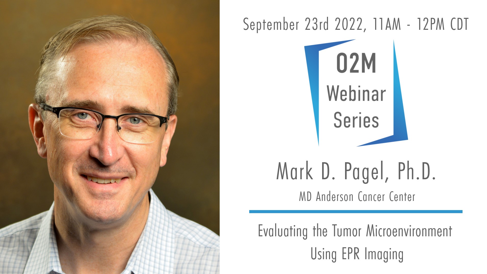
Dr. Marty Pagel has focused on molecular imaging research during the last 20 years in industry and academia. He is a Professor in the Departments of Cancer Systems Imaging and Imaging Physics at the University of Texas MD Anderson Cancer Center. In addition, Dr. Pagel has held leadership positions in professional societies, funding agencies, and scientific journals that focus on molecular imaging. Dr. Pagel’s current research focuses on CEST MRI, PET/MRI, photoacoustic imaging, and EPR imaging, for studies of tumor hypoxia, acidosis, vascular perfusion and enzyme activity, with mouse models of cancer and for clinical trials with cancer patients.
Abstract: Previous studies with EPR imaging have shown that measurements of tumor pO2 can indicate the status of hypoxia in the tumor microenvironment, which can be used to predict response to radiation therapy. To build on these previous studies, we are developing Oxygen Enhanced (OE) EPR imaging that challenges a pre-clinical tumor model with 21% O2 (medical grade air) and 100% O2 in the anesthetic carrier gas, which can be a useful biomarker for evaluating early response to radiation therapy. In addition, we are developing Dynamic Contrast Enhanced (DCE) EPR imaging that measures tumor vascular perfusion, as a complimentary biomarker for evaluating early response to cancer treatment. This presentation will also discuss our ongoing research studies that show how tumor hypoxia causes resistance to immunotherapy, and how reducing hypoxia can improve tumor control with immune checkpoint blockade.
