Webinar

Oxygen Imaging for Efficient Cancer Therapies

This spring, O2M is hosting a webinar mini-series on cancer hypoxia! Join us next month for a conversation with Duke University team Drs. Greg Palmer, Yvone Mowery, & Ashlyn Rickard, investigating how oxygen imaging can optimize the dose and timing of an FDA approved drug papaverine in hypoxia reduction and improved radiation treatment. Then in June hear Dr. Marty Pagel from the University of Texas MD Anderson Cancer Center, exploring tumor microenvironment using oxygen imaging.

Reach out to O2M Technologies to discuss how oxygen imaging can support your cancer research!

May 2022 O2M Webinars

Duke Research Team

Moderator: Prof. Marty Pagel, MD Anderson Cancer Center

About the Speakers:
Greg Palmer is currently an Associate Professor in the Department of Radiation Oncology, Cancer Biology Division at Duke University Medical Center. His primary research focus has been identifying and exploiting the changes in absorption, scattering, and fluorescence properties of tissue associated with cancer progression and therapeutic response.

Yvonne M. Mowery,  MD, PhD, DABR is a physician scientist and the Butler Harris Assistant Professor or Radiation Oncology at Duke University, specializing in treating head and neck cancer with radiotherapy and studying novel combinations of radiation with immunotherapy. Yvonne also serves as the Associate Center Director for Radioimmunotherapy for the Duke Cancer Institute Center of Cancer Immunotherapy. She is the PI of an investigator-initiated phase I trial (NCT04576091) evaluating the ATR inhibitor BAY 1895344 with pembrolizumab and stereotactic body radiation therapy for recurrent HNSCC.

Ashlyn Rickard, PhD, received her doctoral degree in Medical Physics at Duke University in 2022, where she specialized in diagnostic imaging systems and radiation biology. In the Radiation Oncology laboratory of her PhD advisor, Gregory Palmer, PhD, she studies tissue-oxygen-imaging techniques using optical nanoprobes, Cherenkov emission imaging and electron paramagnetic resonance oxygen imaging. Because oxygen imaging has clinical implications in cancer research and radiation biology, most studies included radiation therapy and other anti-cancer therapies.

Abstract: Hypoxia, a prevalent characteristic of most solid, malignant tumors, contributes to diminished therapeutic responses and more aggressive phenotypes. The impact of hypoxia on radiotherapy response is significant: hypoxic tissue is 3x less radiosensitive than normoxic tissue. The major challenge in implementing hypoxic radiosensitizers is the lack of a high-resolution imaging modality that can directly quantify tissue oxygen concentration. A precommercial EPR oxygen-imager was used to quantify tumor hypoxia and investigate the hypoxia-modifying effects of the FDA-approved vasodilator papaverine (PPV). We aimed to quantify the change in absolute tumor hypoxia induced by papaverine in two murine tumor models: E0771 syngeneic mammary carcinoma and primary p53/MCA sarcomas. We hypothesized that 1) there is a PPV dose-related change in tissue pO2, 2) papaverine radiosensitizes tumors, increasing tumor control and survival probability, and 3) pre-screening tumors for baseline tumor hypoxia by EPR imaging predicts radiosensitization in response to PPV. We report that papaverine alters tumor hypoxia in the breast cancer model with an average 47.5% increase in median tumor pO2 and an average 7.8% decrease in tumor hypoxic fraction (<10mmHg); however, no radiosensitizing effect was apparent in either cancer model. We confirmed that hypoxic tumors are more radioresistant than normoxic tumors in the primary sarcoma model (p=0.0057) via oxygen quantification with EPR. Additionally, in a Cox Hazard Regression analysis for the sarcoma model, baseline hypoxic fractions proved to be a significant (p=0.0063) hazard in survivability. Papaverine’s effect on tumor vasculature (in combination with its oxygen consumption rate decrease) requires further study before concluding it is a hypoxic radiosensitizer.

September 2022 O2M Webinar

1e1bb326-f53f-0b03-88d8-a0bc20ae9257

Moderator: Prof. Martyna Elas, Jagiellonian University, Krakaw, Poland

About the Speaker:
Dr. Marty Pagel has focused on molecular imaging research during the last 20 years in industry and academia.  He is a Professor in the Departments of Cancer Systems Imaging and Imaging Physics at the University of Texas MD Anderson Cancer Center.  In addition, Dr. Pagel has held leadership positions in professional societies, funding agencies, and scientific journals that focus on molecular imaging.  Dr. Pagel’s current research focuses on CEST MRI, PET/MRI, photoacoustic imaging, and EPR imaging, for studies of tumor hypoxia, acidosis, vascular perfusion and enzyme activity, with mouse models of cancer and for clinical trials with cancer patients.

Abstract: Previous studies with EPR imaging have shown that measurements of tumor pO2 can indicate the status of hypoxia in the tumor microenvironment, which can be used to predict response to radiation therapy.  To build on these previous studies, we are developing Oxygen Enhanced (OE) EPR imaging that challenges a pre-clinical tumor model with 21% O2 (medical grade air) and 100% O2 in the anesthetic carrier gas, which can be a useful biomarker for evaluating early response to radiation therapy.  In addition, we are developing Dynamic Contrast Enhanced (DCE) EPR imaging that measures tumor vascular perfusion, as a complimentary biomarker for evaluating early response to cancer treatment.  This presentation will also discuss our ongoing research studies that show how tumor hypoxia causes resistance to immunotherapy, and how reducing hypoxia can improve tumor control with immune checkpoint blockade.

Oxygen Imaging for Efficient Cancer Therapies Read More »

Radiation Dose Reduction

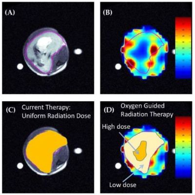
Fibrosarcoma in mouse leg encircled in purple. A) MRI, B) EPROI C) Uniform radiation dose D) Radiation dosed by regional hypoxia
Radiation therapy is a dual-edged sword, capable of destroying cancer cells but also indiscriminately damaging the body in the same process. New research aims to reduce a patient's radiation exposure while still retaining therapeutic efficacy. Tumors are notorious for having a disorganized vascular supply and uneven oxygenation. Regions of tumor that can survive in hypoxic environments are more likely to resist radiation therapy. Understanding the distribution of oxygen may be a key piece of knowledge in achieving that goal. Using Oxygen Guided Radiation Therapy (OGRT), overall radiation dose volume can be reduced without compromising tumor cure.
6035427d-c42b-7479-4572-5dedd07a2a78

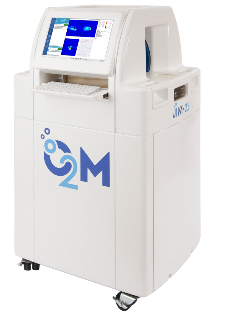
JIVA-25™ is a pre-clinical oxygen imager that provides 3-dimensional oxygen maps of tissues. Learn more about the oxygen imaging technology at www.oxygenimaging.com. Recently, this technology was applied in improving tumor control by localizing tumor radiation to the areas of regional hypoxia. Learn more at our O2M webinar.

76d69d7f-77d6-e469-fa94-a32ecaa75e42

Moderator: Dr. Paul Grippo, University of Illinois at Chicago

About the Speaker:
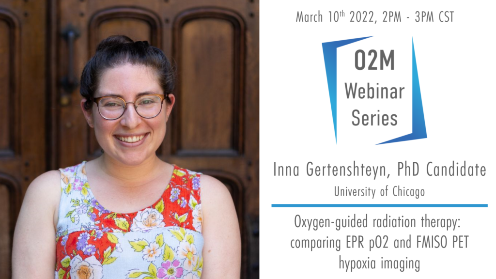
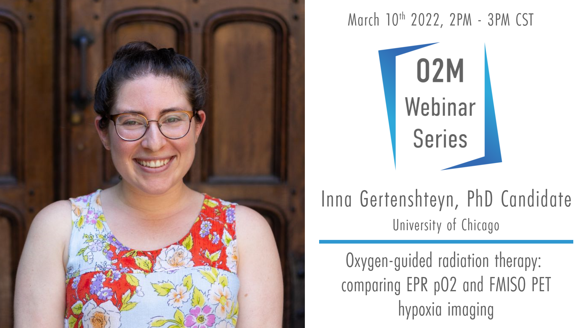
Inna Gertsenshteyn is a PhD candidate at the University of Chicago with co-advisors Dr. Howard Halpern and Dr. Chin-Tu Chen. Her dissertation focuses on multi-modal imaging of tumor hypoxia with EPRI, FMISO PET, and DCE-MRI to improve image-guided radiotherapy. She has won first place at two Young Investigator Award sessions for her work, and is a recipient of the F31 National Research Service Award from the NIH. Before graduate school, Inna was a Senior Image Analyst at Invicro, a global research partner to pharmaceutical and biotech organizations to enhance drug discovery and development. She is currently at Biogen as a Co-op in Late Discovery Imaging focusing on drug development for neuromuscular diseases. Inna plans to graduate in late summer of 2022, and is currently exploring post-doctoral and industry positions.

Abstract:
Hypoxia is a major source of tumor resistance to radiation. Previous studies in the Halpern Lab at the University of Chicago using low-frequency pulsed electron paramagnetic resonance imaging (LF-EPRI) showed significantly improved local tumor control when directing a radiation boost to hypoxic tumor subregions. Those studies were completed in two preclinical tumor types: FSa fibrosarcomas and MCa-4 mammary adenocarcinomas. The presented study was repeated in SCC7 squamous cell carcinomas using both the LF-EPRI system and the higher-frequency JIVA-25™ system from O2M Technologies. Here we will show improved results using the JIVA-25™ for oxygen-guided radiation therapy. We will also compare hypoxia imaging between pO2 EPRI and 18F-Fluoromisonidazole positron emission tomography (FMISO PET), a more clinically-relevant imaging modality, to review how EPRI can help inform the accuracy of PET hypoxia radiotracers with a hybrid PET/EPR imaging system.

Radiation Dose Reduction Read More »

Rapid Scan EPR

956af2e0-ca2e-6d54-2376-1c2dd5b92858

Moderator: Dr. Benoit Driesschaert, West Virginia University

About the Speaker:
Dr. Tseytlin grew up in a family of medical doctors (four generations now) but had a taste for physics, mathematics, and engineering. After receiving his Ph.D. in the field of imaging of conducting materials (physics and mathematics), his interests had shifted towards biology and medicine. He has been developing novel imaging methods with a major focus on biomedical research, as evident from the NIH support. The methods he develops are based on the Electron Paramagnetic Resonance (EPR) phenomenon that was discovered in his alma mater, Kazan State University, in 1944. As a consequence, he had the honor to be taught by the best representatives of the Russian magnetic resonance school. After immigrating to the US, he worked in several leading EPR groups at the University of Denver, University of Chicago, and Geisel School of Medicine at Dartmouth College. In 2015, he was invited to join a newly organized In Vivo Multifunction Magnetic Resonance Center at West Virginia University, where he is now a faculty member (Associate Professor).

Abstract: Rapid scan (RS) EPR is poised to become a mainstream technology given recent developments in hardware, deconvolution methods, and commercialization by the Bruker BioSpin corporation. The RS technique has its constraints and advantages. Some EPR applications, such as functional imaging (including oxygen), will benefit the most. For others, the use of the current state-of-the-art RS EPR method may not be so beneficial. This talk will give an introduction to RS EPR and outline the areas of applications where this method is superior to the standard first-harmonic first-derivative continuous-wave EPR.  In addition, several applications of RS EPR will be demonstrated.


Any researcher reliant on acquisition techniques to obtain their data dreams about having faster acquisition speeds and a high signal-to-noise ratio (SNR). In the context of conventional continuous wave (CW) EPR spectroscopy and imaging, Rapid Scan EPR (RS-EPR) is a revolutionary technique offering exactly that. The short acquisition times, down to milliseconds, open the possibility to observe short-living transient species in chemical reactions or rapidly obtain images. A multifold, and in some cases several hundreds-fold, increase in the SNR allows previously unseen signals to be observed. Due to these advantages, Rapid Scan EPR has quickly gained popularity in the EPR research community.

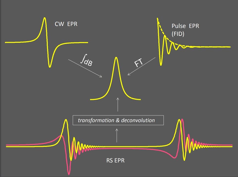
A visual summary of EPR acquisition methods: CW, Pulse, and RS-EPR. Tseytlin, M. Rapid Scan EPR Workshop. 2013(July). DU EPR Center, University of Denver. A visual summary of EPR acquisition methods: CW, Pulse, and RS-EPR. Tseytlin, M. Rapid Scan EPR Workshop. 2013(July). DU EPR Center, University of Denver. CW EPR measures the signal in an equilibrium state with implied limitations on how much power can be delivered, or how fast an acquisition can be performed. In many scenarios, this caps the performance of the instrumentation. RS-EPR is an inherently non-equilibrium technique that can acquire as fast as the instrumentation allows and gain signal from this regime. RS-EPR relies on modern instrumentation such as transient digitizers, high-speed amplifiers, special sweeping coils, and software algorithms for signal reconstruction. All of these are subjects of active research and development. Dr. Mark Tseytlin, Associate Professor in the Department of Biochemistry at West Virginia University, is one of the researchers pushing the boundaries of this novel and exciting technique. Find out more at his recent webinar.

Rapid Scan EPR Read More »

OxyChip Phase 1 Clinical Trial

Moderated by: Dr. Boris Epel, University of Chicago

About the Speaker: Dr. Kuppusamy is a tenured Professor in the Departments of Radiology and Medicine at the Geisel School of Medicine, Dartmouth College. He received his PhD in 1986 from the Indian Institute of Technology, Chennai, India.  Following a 2-year Fogarty Fellowship from NIH/NIA, he became a faculty in the Department of Medicine at the Johns Hopkins University School of Medicine, in 1987. He then moved to the Ohio State University in 2002, where he was holding the William D. and Jacquelyn L. Wells Chair in Imaging Research. He also served as the Associate Director of the Division of Cardiovascular Medicine. He joined the Geisel School of Medicine at Dartmouth College in 2013. Dr. Kuppusamy’s research interests include free radicals, oxygen and oxygen biology in cardiovascular diseases and cancer. He is well-known for his expertise in the development of imaging methods for imaging oxygen and free radicals in biological systems. Dr. Kuppusamy has received numerous research awards including Silver Medal in 2006 from the International EPR Society for significant contribution to the development of EPR imaging for biomedical applications and Doctor of Medicine (honoris causa) in 2008 from University of Pecs (Pecs, Hungary) for cardiovascular research. Dr. Kuppusamy has published 400 peer-reviewed research manuscripts in leading biomedical journals. He has received several research grants from the American Heart Association and National Institutes of Health. He has several patents on the probes and methods for determination of oxygen concentration in tissues. One of his inventions, OxyChip, is currently in a Phase I clinical trial.

About the Webinar: Clinical interventions to mitigate the impact of tumor hypoxia on cancer-treatment outcomes have been hampered by an inability to assess patient-specific tumor oxygen levels. The overall objective of this first-in-human study was to assess the safety and feasibility a novel oxygen sensor, called ‘OxyChip’, as a clinically viable technology to make individualized tissue-oxygen assessments. OxyChip is a paramagnetic oxygen sensor composed of oxygen-sensing lithium octa-n-butoxynaphthalocyanine (LiNc-BuO) crystals embedded in a biocompatible polymer. Patients with any tumor at ≤3-cm depth from the skin surface and scheduled to receive surgical resection (with or without neoadjuvant therapy) were considered eligible. OxyChips were implanted in the tumor and subsequently removed during surgery. Oxygen (pO2) at the sensor location was assessed using electron paramagnetic resonance (EPR) oximetry. During each measurement session, the patient breathed room air, followed by a period of oxygen inhalation using a non-rebreather mask with 100% oxygen delivered at a flow rate of 15 l/min, and then breathed room air again. Twenty-four patients underwent OxyChip implantation in their tumors. Six patients received neoadjuvant therapy. Median implant duration was 29 days (range 4–128 days). The OxyChips recovered from the patients did not show any significant change in their oxygen sensitivity. The implantation procedure and the process of EPR oximetry in the clinic were well tolerated by patients. Histopathologic findings revealed no clinically significant pathology, indicating that the tissue reaction to the OxyChip was well within expectations for an implanted device [1]. A total of 44 measurements were made in 15 patients. Baseline tumor pO2 was variable with overall median of 10.2 mmHg (range 0–48.5 mmHg); 48% of the values were below clinically significant hypoxia (10 mmHg). After hyperoxygenation, the overall median pO2 was 21.4 mmHg (range 0.4–97.6 mmHg). For 61% of measurements, there was a statistically significant (p<0.05) response to hyperoxygenation. Measurement of baseline pO2 and response to hyperoxygenation using EPR oximetry with OxyChip is clinically feasible in a variety of tumor types. Tumor oxygenation at baseline also differed significantly among patients, with about half exhibiting clinically significant hypoxia at baseline. Although most tumors responded to hyperoxygenation intervention, some were non-responders. The pO2 data demonstrated the need for individualized assessment of tumor oxygenation in the context of planned hyperoxygenation interventions to optimize clinical outcomes.

References:
[1] Schaner PE, Pettus JR, Flood AB, Williams BB, Jarvis LA, Chen EY, Pastel DA, Zuurbier RA, diFlorio-Alexander RM, Swartz HM, Kuppusamy P. OxyChip implantation and subsequent EPR oximetry in human tumors is safe and feasible: First experience in 24 patients. Front Oncol 10:572060 (2020).

OxyChip Phase 1 Clinical Trial Read More »

Rapid Scan EPR of Nitroxide Probes of Redox

Moderated by: Dr. Benoit Driesschaert, West Virginia University

About the Speaker:Gareth R. Eaton received his A.B. at Harvard and obtained his Ph.D. at MIT 1972. Since then he has been in the Department of Chemistry (now the Department of Chemistry and Biochemistry) at the University of Denver. He was promoted to Professor in 1980. He served as Dean of Natural Sciences from 1984-1988 and as Vice Provost for Research from 1988-1989.  In 1997 he received the John Evans Professorship at the University of Denver. He teaches inorganic chemistry. His research program involves continuous wave, rapid-scan, and pulse EPR applied to the study of relaxation times, spin-spin interaction, metal ions in biological systems, and EPR imaging. He and his wife, Professor Sandra S. Eaton, have jointly authored over 400 research papers and book chapters. In 2002 they jointly received the Bruker prize and in 2008 they became Fellows of the International EPR/ESR Society. He is a Fellow of the Royal Society of Chemistry.

About the Webinar: A brief introduction to the relative benefits of CW, rapid scan, and pulsed EPR for solving various problems will be presented.  Emphasis will be on applications to nitroxide probes designed to measure redox status of tissues.

Rapid Scan EPR of Nitroxide Probes of Redox Read More »

EPR Imaging Outside the Pre-Clinic

Moderated by: Dr. Howard Halpern, University of Chicago

About the Speaker: Dr. Biller is a Principal Chemist at TDA Research, Inc. in Golden, CO. He graduated from the Eaton Laboratory at the University of Denver in 2014 and was awarded the Bruker Biospin/Royal Society of Chemistry International Thesis award in 2015. After graduation, Dr. Biller went to work with the Magnetic Imaging Group at NIST-Boulder, first as a National Research Council Post-doctoral scholar, and then as a Research Chemist. In 2018 he joined TDA Research and has driven the development of novel non-destructive evaluation techniques based on the interaction of electromagnetic waves with materials including carbon fiber and lithium-ion batteries. Dr. Biller has a deep interest in widening the application sphere of magnetometry, electron paramagnetic resonance, and nuclear magnetic resonance methods outside of the laboratory environment. He is currently leading an NCI SBIR Phase I project to develop an in-vivo EPR imaging agent for clinical use.

About the Webinar: EPR Imaging is a powerful technique in pre-clinical small animal applications. Through EPRI, a spatial map of oxygen concentration can be created and used to guide targeted increased doses of radiation at hypoxic sites. This has been shown to improve the survivability of a mouse model. The transition of this very useful new tool into the clinic has not been straightforward. Concerns about toxicity of the imaging agents, a lack of familiarity with the EPR imaging hardware required, and the way in which the EPRI agent would be administered are all hurdles. This presentation will touch on the development of a novel EPRI imaging agent where the paramagnetic probe has been attached to a nanoparticle and encapsulated with an oxygen-permeable membrane. This separates the body and imaging agent environment, while still allowing O2 to access the paramagnetic site. This work, led by TDA Research Inc., has been supported by collaborations with West Virginia University, The University of Denver, and O2M Technologies under an SBIR Phase I administered by the National Cancer Institute (NCI Contract #75N91020C00032).

EPR Imaging Outside the Pre-Clinic Read More »

Development of Stable Triarylmethyl Radicals for Biomedical EPR Applications

About the Speaker: Dr. Benoit Driesschaert is an Organic Chemist, Assistant Professor at West Virginia University in the Department of Pharmaceutical Sciences. His laboratory develops stable triarylmethyl radicals used as spin probes and spin labels for biomedical EPR applications.​

About the Webinar: Low-field electron paramagnetic resonance (EPR) with a molecular spin probe is a powerful technique to profile various biomarkers of the tissue microenvironment in vivo. This presentation will discuss the design, synthesis, and applications of triarylmethyl (TAM) radicals sensitive to important physiological parameters such as dissolved oxygen concentration, pH, inorganic phosphate (Pi) concentration, microviscosity, and enzyme activity.

Development of Stable Triarylmethyl Radicals for Biomedical EPR Applications Read More »

In Vitro EPR Spectroscopy: A Complementary Technique with EPR Imaging

About the Speaker: Harold Swartz MD, PhD, MSPH, has held the Alma Hass Milham Distinguished Chair in Clinical Medicine and has been the director of the Electron Paramagnetic Resonance (EPR) Centers at Dartmouth, the University of Illinois and the Medical College of Wisconsin. His current interests are focused especially on the measurement of oxygen in vivo to improve diagnosis and therapy in the wide range of pathophysiology for which tissue oxygen is a critical parameter. He has especially been involved in the development of EPR to make measurements in human subjects to facilitate measurements of oxygen and radiation exposure. He has authored/co-authored over 550 publications and six books. His honors include the International Zavoisky Award (2005); Fellow of International EPR (ESR) Society (2005); special gold medal as Founder of the International EPR Society (2005); Fellow of International Society for Magnetic Resonance in Medicine (1997); silver medal (Biomedicine) from International EPR Society (1994), and the silver medal from the Society for Magnetic Resonance in Medicine (1993).

About the Webinar: EPR spectroscopy provides quantitative measurement of oxygen based on the physical interaction of molecular oxygen with the paramagnetic materials, modifying the EPR spectra proportionally. Two different types of paramagnetic materials can be used for EPR spectroscopy: soluble free radicals (such as nitroxides and trityls) and particulates such lithium phthalocyanine and carbon particles. The particulates may higher spin densities. The soluble free radicals may be especially valuable for oximetry because they can have especially valuable because in addition to measuring oxygen, they also can measure a number of other biochemical and physical parameters within the capsule such as pH, viscosity, and SH groups. In my webinar, I will cover applications of EPR spectroscopy to various applications.

In Vitro EPR Spectroscopy: A Complementary Technique with EPR Imaging Read More »

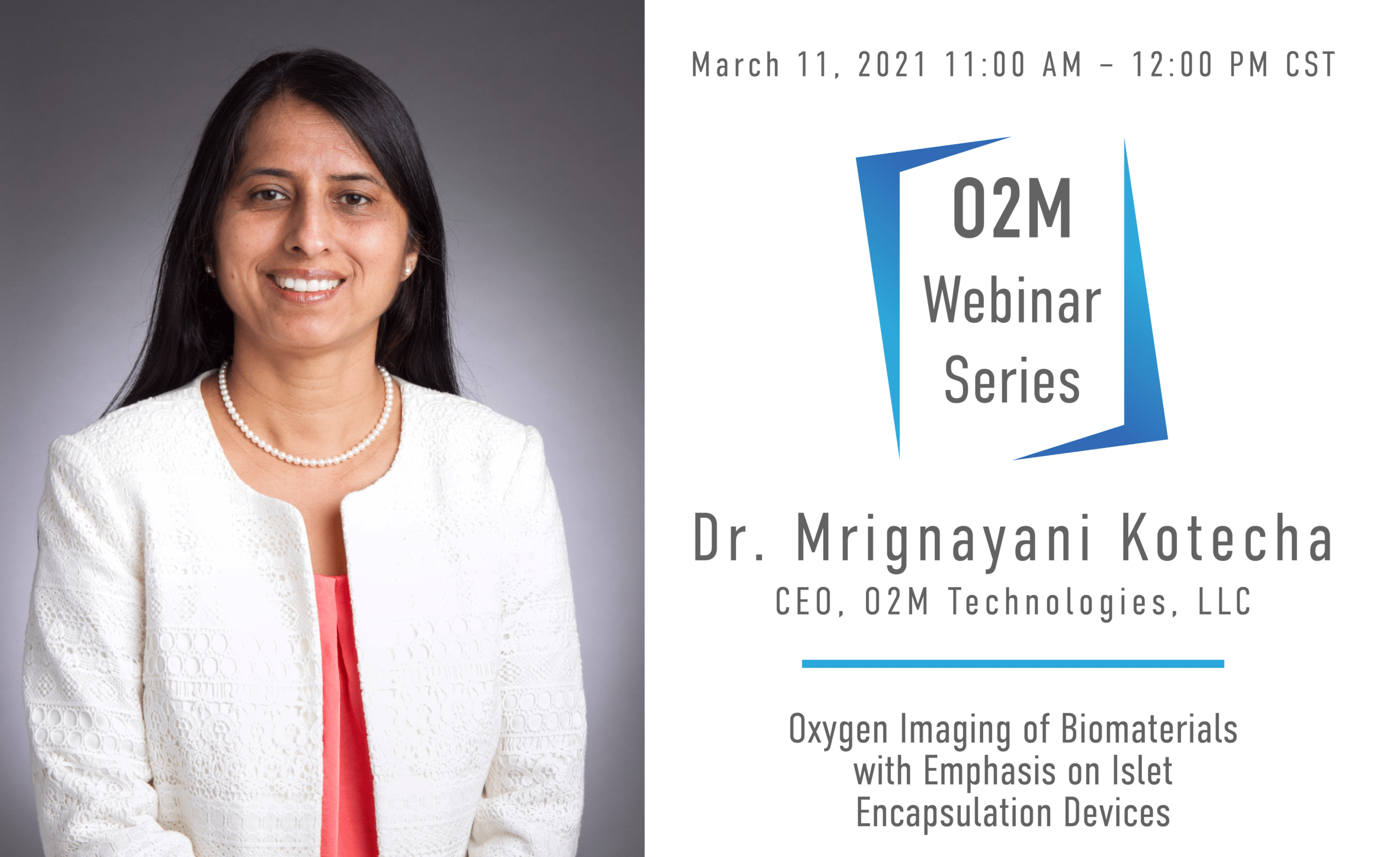
Oxygen Imaging of Biomaterials with Emphasis on Islet Encapsulation Devices

2fa973c1-3ecc-4c86-8cca-2236810091db

Moderated by: Carl G. Simon, Jr., Ph.D., National Institute of Standards and Technology (NIST)
About the Speaker: Dr. Kotecha is a founding member and CEO of O2M technologies. She graduated from the Physics program from Jabalpur University, India in solid state NMR of quadrupolar nuclei in 2001. During her academic career before O2M, she has worked as a full-time tenured professor of Physics at Government Science College, Jabalpur; as a visiting scientist at Indian Institute of Science, Bangalore, at Weizmann Institute of Science, University of Duisburg-Essen, and University of Illinois at Chicago. In 2017, she co-founded O2M Technologies to bring the EPR oxygen imaging technology to the market. Dr. Kotecha is an expert in magnetic resonance methodology, instrumentation, pulse sequence design, and applications of MRS/MRI to biomaterials, engineered tissues, nanoparticles, membrane proteins, cryolites, ferroelectrics, and cancer. She is an author of more than 40 publication and the lead editor of 2017 Wiley book “Magnetic Resonance in Tissue Engineering”. She developed an ASTM International standard F3224-17 “Standard Test Method for Evaluating Growth of Engineered Cartilage Tissue using Magnetic Resonance Imaging” that received CDRH recognition in 2019.

About the Webinar: The lack of oxygen supply to the highly metabolic pancreatic islet cells is one of the major factors contributing to the failure of islet transplantation devices targeting the cure of type I diabetes (T1D). The loss of islets due to hypoxia is common in almost all modes of islet transplantation- micro-encapsulation devices, macro-encapsulation devices, and tissue-grafts transplantation. Several approaches to improve oxygenation in these transplantation devices are thus being tested. However, because of the lack of available technologies to provide oxygen partial pressure (pO2) assessment in and around devices, the progress is severely hindered.
O2M Technologies’ platform non-invasive Oxygen Imaging technology has the potential to guide the development of islet cell transplantation therapies by providing real-time high accuracy pre- and post-implantation pO2 maps in and around devices in vitro and in vivo. O2M’s preclinical small animal oxygen imager, JIVA-25, provides average pO2 values in sample volumes (up to 40 mm) as well as three-dimensional pO2 maps with high spatial (0.5 mm, isotropic), temporal (1-10 min), and pO2 (1-3 torr) resolution. For reporting oxygen concentration, JIVA-25 uses oxygen-dependent relaxation rates of trityl radicals OX063 or its deuterated version OX071. This work is the outcome of JDRF-supported “Oxygen Measurement Core” facility established at O2M Technologies in 2019. We performed in vitro and in vivo pO2 measurements of acellular and cell loaded islet cell transplantation devices. These devices vary in shape, size, biomaterials, and oxygen profile. I will present the key data from these measurements in my webinar.

Oxygen Imaging of Biomaterials with Emphasis on Islet Encapsulation Devices Read More »

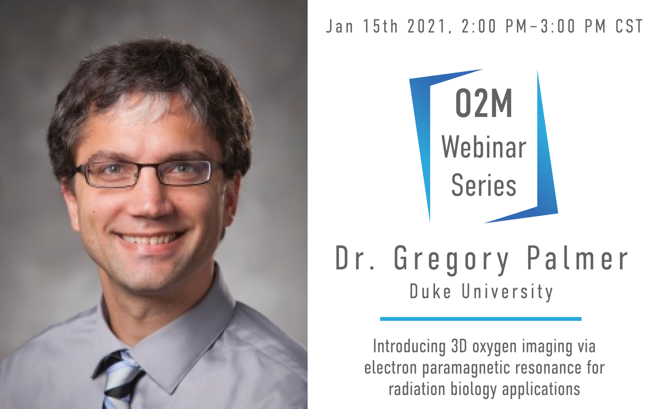
Introducing 3D Oxygen Imaging VIA EPR for Radiation Biology Applications

2f9ade26-0c64-4421-954a-a01a874df253

Moderated By: Dr. Mark Dewhirst, Duke University

About the Speaker: Greg Palmer obtained his B.S. in Biomedical Engineering from Marquette University in 2000, after which he obtained his Ph.D. in BME from the University of Wisconsin, Madison. He is currently an Associate Professor in the Department of Radiation Oncology, Cancer Biology Division at Duke University Medical Center. His primary research focus has been identifying and exploiting the changes in absorption, scattering, and fluorescence properties of tissue associated with cancer progression and therapeutic response. To this end he has implemented a model-based approach for extracting absorber and scatterer properties from diffuse reflectance and fluorescence measurements. More recently he has developed quantitative imaging methodologies for intravital microscopy to characterize tumor functional and molecular response to radiation and chemotherapy. His awards have included the Jack Fowler Award from the Radiation Research Society.

About the Webinar: Hypoxia severely limits the efficacy of radiotherapy, chemotherapy, and immunotherapy. This results in diminished success in treating cancer. Despite consistent research in this field and treatments with promising anti-hypoxia mechanisms, there is no treatment for ameliorating hypoxia. This could be a result of ineffective hypoxia mitigators or because there is not a clinical imaging modality capable of directly quantifying oxygen at high temporal, spatial and oxygen resolution. Electron paramagnetic resonance oxygen imaging (EPROI) offers 3D oxygen maps at high resolution. The first, commercial, preclinical EPROI unit, JIVA-25 developed by O2M, is undergoing its final validation. At Duke University, this EPROI system is currently being utilized in radiation biology applications, investigating a promising radiation sensitizer in the E0771 murine flank tumor model. Oxygen microbubbles are venously-introduced and burst in the tumor via ultrasound, providing a bolus of oxygen. Preliminary in vitro data reports that an increase in tumor pO2 mere milliseconds before radiation significantly increases radiation-induced cancer cell damage. In vivo pilot studies have shown an increase in hemoglobin saturation in murine tumor models; however, to fully elucidate the role of oxygen microbubbles in alleviating hypoxia and to determine the ideal timing of prospective radiotherapy, the increase in tissue pO2 must be directly quantified spatially and temporally. We hypothesize that oxygen microbubbles will cause an acute increase in tumor pO2. Here, we present our initial data from this study. These described experiments, while focusing on the radiation biology field, will provide a preclinical imaging paradigm for quantifying hypoxia in vivo that is applicable to any research that requires precise evidence of tissue pO2.

Introducing 3D Oxygen Imaging VIA EPR for Radiation Biology Applications Read More »

Scroll to Top