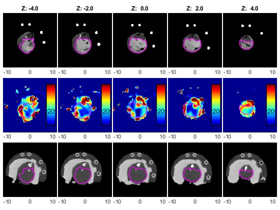
Tissue oxygenation is determined by the balance of oxygen delivery to tissues against oxygen consumption by those tissues. Normally, this is a well-maintained balance through several homeostatic mechanisms. However, solid tumors are unable to maintain oxygen balance due to the aberrant structure and function of a tumor’s vascular supply, the tumor microenvironment, and the intense metabolic demands of tumor cells. In some cases, tumor cells develop adaptive strategies to escape oxygen deficiency, whereby hypoxic environments act as a selective pressure resulting in clonogenic expansion of tumor cells with hypoxia tolerance. This establishes a vicious cycle of hypoxia and malignant progression. Moreover, hypoxic tumor environments reduce the effectiveness of radiation therapy, as oxygen needs to be present within milliseconds of radiation to observe the “oxygen effect” - whereby oxygen radicals enhance radiation induced DNA damage. It has been well demonstrated in sarcomas of the cervix, breast, and prostate that hypoxia can be considered an independent predictor of disease progression, treatment failure, and metastatic potential. Electron paramagnetic resonance oxygen imaging is a technology 20 years in the making, that can non-invasively capture real time absolute-value images of oxygen tension in solid tumors (see figure).
Learn more about tumor oximetry at Dr. Martyna Elas's webinar.
