Webinar

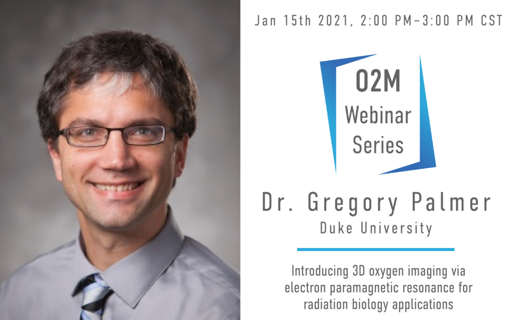
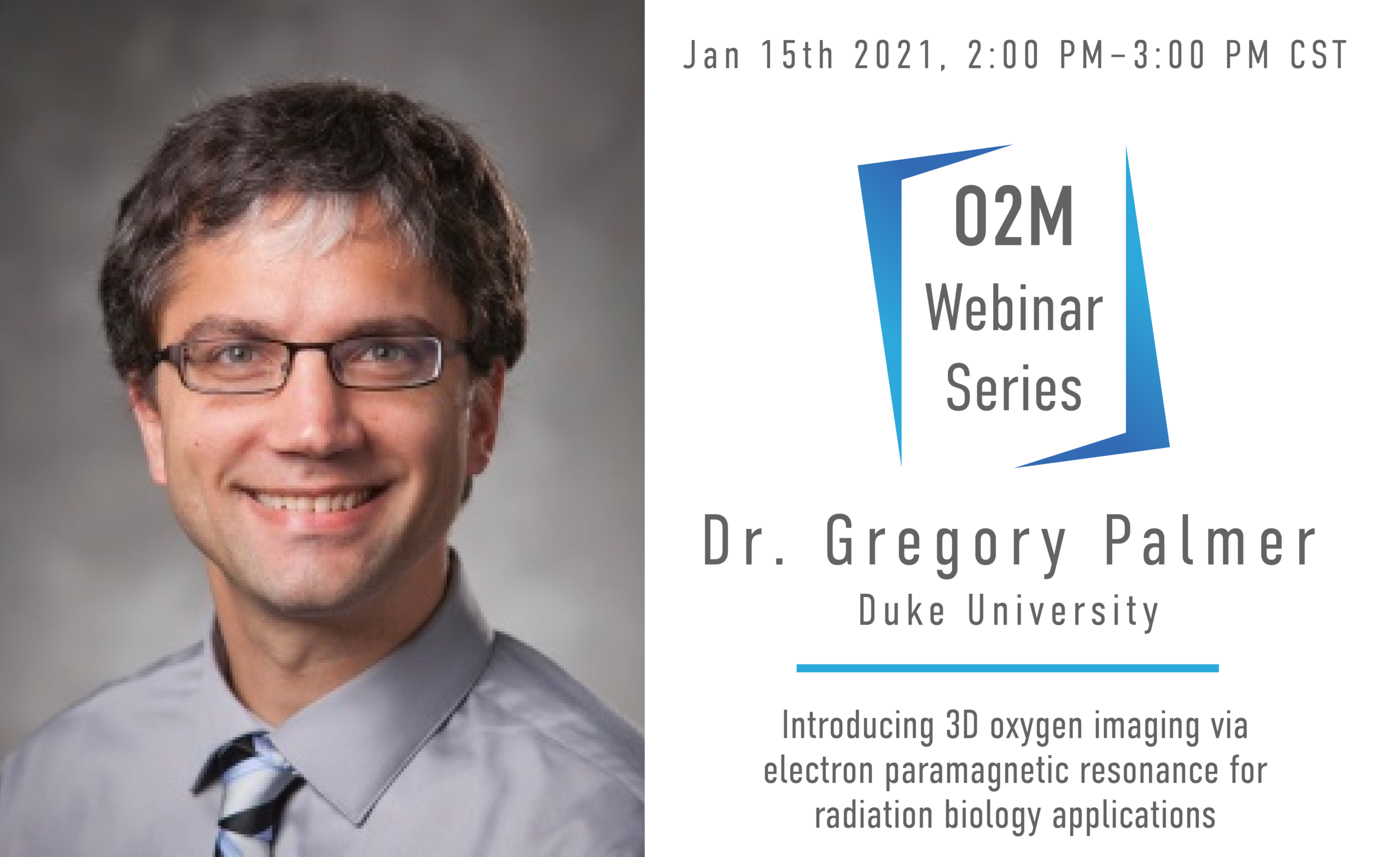
Introducing 3D Oxygen Imaging VIA EPR for Radiation Biology Applications
2f9ade26-0c64-4421-954a-a01a874df253

Moderated By: Dr. Mark Dewhirst, Duke University

About the Speaker: Greg Palmer obtained his B.S. in Biomedical Engineering from Marquette University in 2000, after which he obtained his Ph.D. in BME from the University of Wisconsin, Madison. He is currently an Associate Professor in the Department of Radiation Oncology, Cancer Biology Division at Duke University Medical Center. His primary research focus has been identifying and exploiting the changes in absorption, scattering, and fluorescence properties of tissue associated with cancer progression and therapeutic response. To this end he has implemented a model-based approach for extracting absorber and scatterer properties from diffuse reflectance and fluorescence measurements. More recently he has developed quantitative imaging methodologies for intravital microscopy to characterize tumor functional and molecular response to radiation and chemotherapy. His awards have included the Jack Fowler Award from the Radiation Research Society.

About the Webinar: Hypoxia severely limits the efficacy of radiotherapy, chemotherapy, and immunotherapy. This results in diminished success in treating cancer. Despite consistent research in this field and treatments with promising anti-hypoxia mechanisms, there is no treatment for ameliorating hypoxia. This could be a result of ineffective hypoxia mitigators or because there is not a clinical imaging modality capable of directly quantifying oxygen at high temporal, spatial and oxygen resolution. Electron paramagnetic resonance oxygen imaging (EPROI) offers 3D oxygen maps at high resolution. The first, commercial, preclinical EPROI unit, JIVA-25 developed by O2M, is undergoing its final validation. At Duke University, this EPROI system is currently being utilized in radiation biology applications, investigating a promising radiation sensitizer in the E0771 murine flank tumor model. Oxygen microbubbles are venously-introduced and burst in the tumor via ultrasound, providing a bolus of oxygen. Preliminary in vitro data reports that an increase in tumor pO2 mere milliseconds before radiation significantly increases radiation-induced cancer cell damage. In vivo pilot studies have shown an increase in hemoglobin saturation in murine tumor models; however, to fully elucidate the role of oxygen microbubbles in alleviating hypoxia and to determine the ideal timing of prospective radiotherapy, the increase in tissue pO2 must be directly quantified spatially and temporally. We hypothesize that oxygen microbubbles will cause an acute increase in tumor pO2. Here, we present our initial data from this study. These described experiments, while focusing on the radiation biology field, will provide a preclinical imaging paradigm for quantifying hypoxia in vivo that is applicable to any research that requires precise evidence of tissue pO2.

Introducing 3D Oxygen Imaging VIA EPR for Radiation Biology Applications Read More »

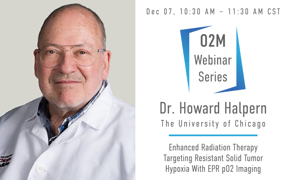
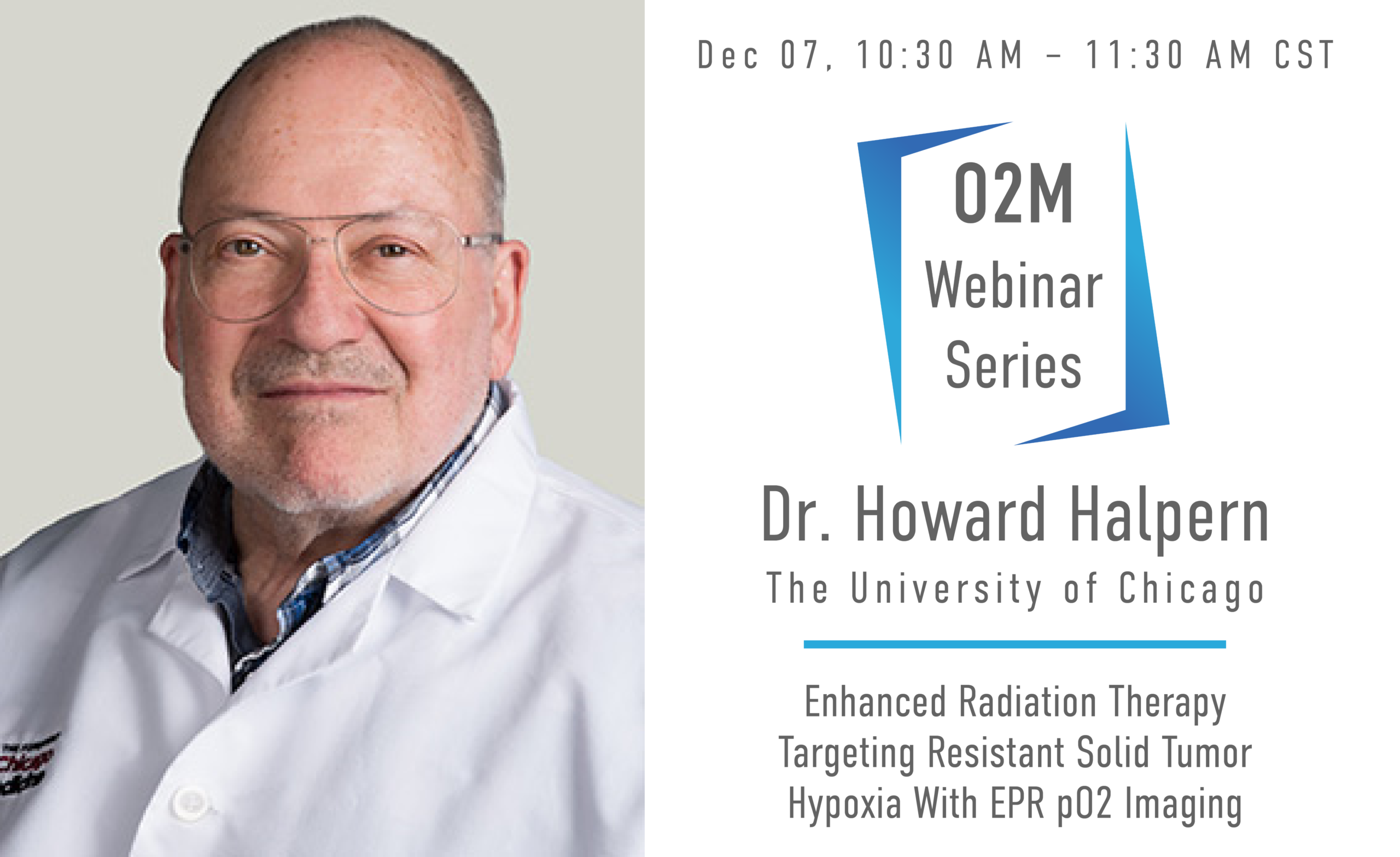
Enhanced Radiation Therapy Targeting Resistant Solid tumor Hypoxia with EPR pO2 Imaging

About the Speaker: Dr. Halpern is a Professor of Radiation and Cellular Oncology and Director (PI) of the NIH Center for EPR Imaging In Vivo Physiology at the University of Chicago and an attending Radiation Oncologist practicing in the University of Illinois clinic run by the University of Chicago. A major focus of his research effort has been to develop high resolution noninvasive images of oxygen distributions in tumors and normal tissues living animals with electron paramagnetic resonance (EPR) imaging.

About the Webinar: Resistant localized portions of solid tumors has been postulated to exist for over six decades.  These have primarily been associated with regions of tumor hypoxia with low molecular oxygen or low pO2.  It was one of the driving concepts behind the development of Intensity Modulated Radiation Therapy (IMRT), where radiation dose could be sculpted to produce large dose gradients within the tumor boundaries.   Positron Emission Tomography, PET, using 18F labeled nitroimizole radiotracers which have been shown to be retained in hypoxic tumor regions was hoped to provide a reliable means to localized resistant hypoxic tumor but this has failed to show benefit.  Quantitative electron paramagnetic resonance (EPR) pO2 images has provided the first demonstration of local tumor control enhancement through identification of tumor hypoxia and directing extra radiation to those hypoxic tumor regions.   We will describe the nature of this magnetic resonance imaging modality using electron resonance rather than nuclear resonance.  A nontoxic spin probe is necessary to report the local pO2.  Low magnetic field strengths, a few hundred times that of the earth's magnetic field prepares the images.  Data from two mammalian tumors, the first in over a century of the observation of hypoxic resistance to radiation will be shown.

Enhanced Radiation Therapy Targeting Resistant Solid tumor Hypoxia with EPR pO2 Imaging Read More »

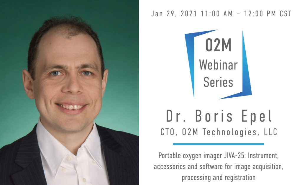
Electron Paramagnetic Resonance Oxygen Imaging, Principles and Applications

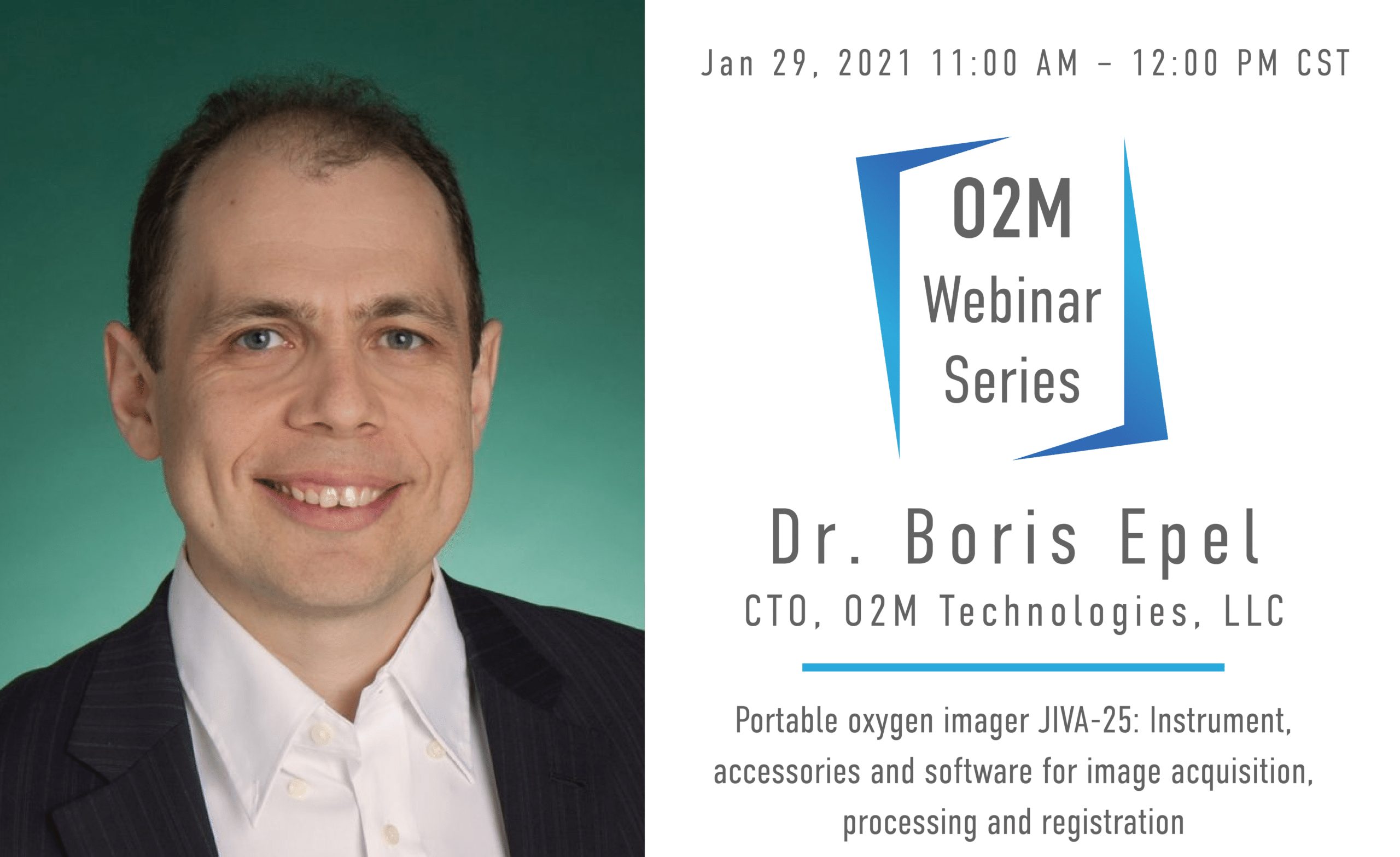
Dr. Boris Epel, CTO, O2M Technologies

About the Speaker: Our first speaker is Dr. Boris Epel, Research Professor of the Department of Radiation and Cellular Oncology, University of Chicago. Dr. Epel is one of the inventors of oxygen imaging technology, the designer of imaging instrument, and a founding member of the company.

About the Webinar: Electron Paramagnetic Resonance (EPR) Imaging is a well-established method for the study of spatial distribution and local environment of electron paramagnetic centers and spin probes. One of the most important applications of modern EPR imaging is in vivo oximetry in which soluble spin probes with oxygen-dependent relaxation rates are used. The webinar will present fundamental principles of EPR oxygen imaging and give an overview of main applications.

Electron Paramagnetic Resonance Oxygen Imaging, Principles and Applications Read More »

Scroll to Top