Newsletters

A Lesson From Insects on Overcoming Cell Encapsulation Hypoxia

Inadequate oxygenation is a major challenge in cell encapsulation, a therapy which holds potential to treat many diseases including type I diabetes. In such systems, cellular oxygen (O2) delivery is limited to slow passive diffusion from transplantation sites through a poorly O2-soluble encapsulating matrix, usually a hydrogel. This constrains the maximum permitted distance between encapsulated cells and their host site to within a few hundred micrometers to ensure cellular function.

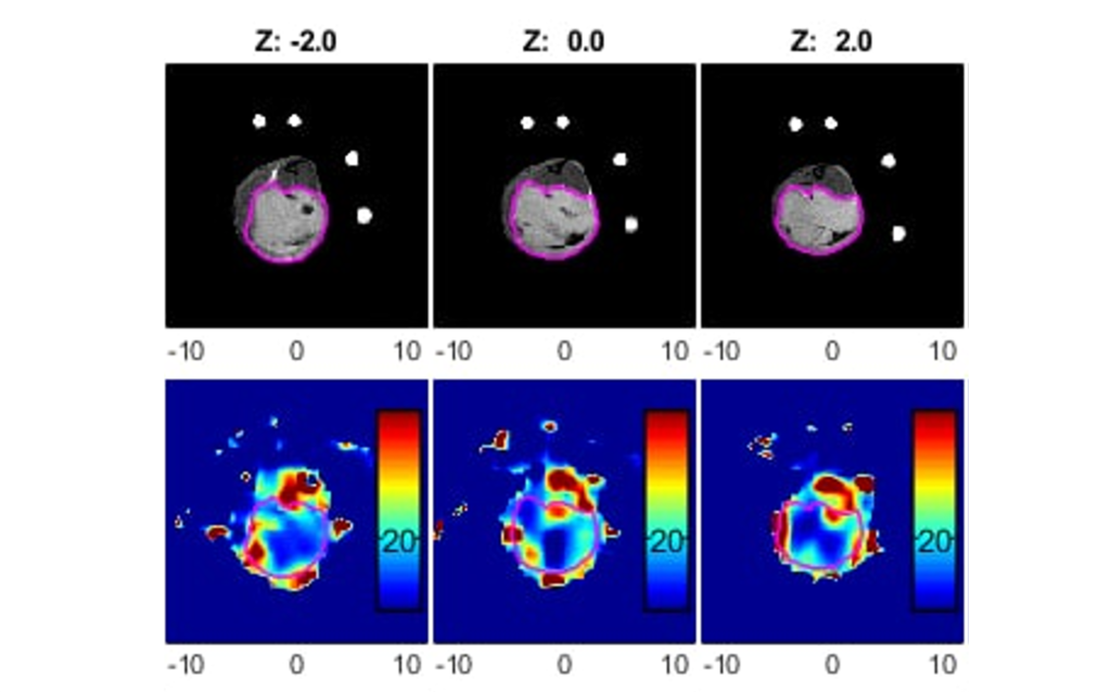
To solve the problem of poor oxygen penetration, the Dr. Minglin Ma's group at Cornell University took inspiration from mealworm beetle larvae! As opposed to the blood circulatory system of vertebrates, many insects transport oxygen through a tracheal system (Fig. 1): a gas-filled, ladder-like channel network that permits O2 distribution across multi-millimeter scales. Critically significant to this design, gaseous O2 has a 104 times higher diffusion coefficient than dissolved O2. Ma's group designed a biomimetic scaffold featuring internal continuous air channels endowed with 10,000-fold higher O2 diffusivity than hydrogels (Fig. 2). They named the scaffold SONIC (Speedy Oxygenation Network for Islet Constructs), reported in last month's Nature Communications.

Figure 1: Beetle larva tracheal system. Figure 2: 3D mold cast for SONIC device
Figure 1: Mealworm beetle larva tracheal system. Figure 2: 3D cast of SONIC device.

The SONIC scaffold is comprised of a hydrophobic polymer (vinylidene fluoride-co-hexafluoropropylene), and the internal continuous air channels were created by a phase separation process in a 3D printed mold. A hydrophilic polydopamine coating was applied to the scaffold surface, providing a compatible interface between the hydrophobic scaffold and hydrophilic hydrogel. SONIC's internal hydrophobicity avoids water penetration into the air channels, an essential feature for enabling high O2 permeability. Finally, a cell-laden hydrogel was applied via a simple in situ cross-linking procedure by pre-deposited CaSO4 crystals on the scaffold surface.

pO2 map and average pO2 of SONIC and control devices

A spiral SONIC device was designed for delivering a clinically relevant islet dose for islet replacement therapy, which computational models predict could support a curative islet dose of 500 k IEQ human islets within a disk approximately 11 cm in diameter.

In summary, the SONIC scaffold provides a solution to the poor transport of O2 in traditionally employed bulk hydrogels of cell encapsulation systems and represents a promising platform for translatable encapsulation devices requiring high cell payloads.

Reach out to O2M™ for your experiments. We provide high resolution oxygen maps for in vitro and in vivo samples, among many other Core services.

A Lesson From Insects on Overcoming Cell Encapsulation Hypoxia Read More »

Tumor Hypoxia Imaging

Axial slices of SCC7 squamous cell carcinoma in mouse leg of MRI, PO2 by JIVA-25™, and CT. Tumor contoured in pink.
Photo credit: Howard Halpern Lab, The University of Chicago
Axial slices of SCC7 squamous cell carcinoma in mouse leg of MRI, PO2 by JIVA-25™, and CT. Tumor contoured in pink. Photo credit: Howard Halpern Lab, The University of Chicago

Tissue oxygenation is determined by the balance of oxygen delivery to tissues against oxygen consumption by those tissues. Normally, this is a well-maintained balance through several homeostatic mechanisms. However, solid tumors are unable to maintain oxygen balance due to the aberrant structure and function of a tumor’s vascular supply, the tumor microenvironment, and the intense metabolic demands of tumor cells. In some cases, tumor cells develop adaptive strategies to escape oxygen deficiency, whereby hypoxic environments act as a selective pressure resulting in clonogenic expansion of tumor cells with hypoxia tolerance. This establishes a vicious cycle of hypoxia and malignant progression. Moreover, hypoxic tumor environments reduce the effectiveness of radiation therapy, as oxygen needs to be present within milliseconds of radiation to observe the “oxygen effect” - whereby oxygen radicals enhance radiation induced DNA damage. It has been well demonstrated in sarcomas of the cervix, breast, and prostate that hypoxia can be considered an independent predictor of disease progression, treatment failure, and metastatic potential. Electron paramagnetic resonance oxygen imaging is a technology 20 years in the making, that can non-invasively capture real time absolute-value images of oxygen tension in solid tumors (see figure).

Learn more about tumor oximetry at Dr. Martyna Elas's webinar.

Tumor Hypoxia Imaging Read More »

The First Publication from “Oxygen Measurement Core”

Cell encapsulation represents a promising therapeutic strategy for many hormone-deficient diseases such as type I diabetes (T1D). However, adequate oxygenation of the encapsulated cells remains a challenge, especially in the poorly oxygenated subcutaneous site.

Recently, our collaborator, Dr. Minglin Ma's group at Cornell University, published a paper “An inverse-breathing encapsulation system for cell delivery” in Science Advances. This work reported a novel oxygen delivery system that generates oxygen (O2) for the islet cells from their own waste product, carbon dioxide (CO2), in a self-regulated (i.e. “inverse breathing”) way, supporting the long-term function of islets in the cell encapsulation device.

As lead author, Dr. Longhai Wang designed and fabricated the inverse-breathing device (iBED) by employing a gas-solid (CO2-lithium peroxide) reaction that was completely separated from the aqueous cellular environment by a gas-permeable membrane (Fig. a and b). Doctoral student Alexander Ernst performed computational modeling which guided device design optimizations (Fig. c).

503544a8-3cab-6a65-eee0-dd4b9d151fef

Oxygen mapping was performed using O2M's preclinical oxygen imager JIVA-25™ instrument at O2M's “Oxygen Measurement Core” facility to validate the CO2-regulated O2 release in the inverse-breathing devices (Fig. d).

14c40f4b-fdce-79ec-44df-17f36a2c00ea

The iBED restored normoglycemia of immunocompetent diabetic mice for over 3 months. And functional islets were observed in scaled-up device implants in minipigs retrieved after 2 months (Fig. e). Furthermore, O2 supply may be extended indefinitely by the introduction of a tank replacement or formulation refilling modular design.

d464d3e5-683a-7397-f703-81ffb5c380f5

This inverse breathing device provides a potential system to support long-term cell function in the clinically attractive subcutaneous site by overcoming several outstanding challenges in oxygenating encapsulated cells and thus represents considerable progress in the use of translatable long-term O2-supplementing technologies for cell replacement therapies.

Read the complete article here: https://advances.sciencemag.org/content/7/20/eabd5835

The First Publication from “Oxygen Measurement Core” Read More »

First Publication from “Oxygen Measurement Core”

81cea4a9-2bca-d625-a788-05739721803c

Did you know that JIVA-25™ can provide in vivo partial oxygen pressure (pO2) maps of an implanted devices in a rodent? The pO2 maps provide a crucial missing information to assess the success of all transplanted devices. Reach out to us if you are interested in performing such study in collaboration with us at our "Oxygen Measurement Core" facility or at your lab by bringing a JIVA-25™ to your lab. Looking forward to talk to you about what excites you in your journey of developing new cell- and tissue-based therapies!

First Publication from “Oxygen Measurement Core” Read More »

In Vivo Oxygen Imaging of Islet Encapsulation Devices using JIVA-25™

81cea4a9-2bca-d625-a788-05739721803c

Did you know that JIVA-25™ can provide in vivo partial oxygen pressure (pO2) maps of an implanted devices in a rodent? The pO2 maps provide a crucial missing information to assess the success of all transplanted devices. Reach out to us if you are interested in performing such study in collaboration with us at our "Oxygen Measurement Core" facility or at your lab by bringing a JIVA-25™ to your lab. Looking forward to talk to you about what excites you in your journey of developing new cell- and tissue-based therapies!

In Vivo Oxygen Imaging of Islet Encapsulation Devices using JIVA-25™ Read More »

O2M at the “Society for Biomaterials 2021 Annual Meeting”

0146dde7-8717-4172-93fb-5ccc08153774

Rapid Fire Presentations

558: In Vitro pO2 Measurement of Islet Encapsulation Devices in Oxygen Measurement Core 
(In collaboration with the University of Chicago, University of Arizona, Cornell University, University of Florida, and McGill University)

This work is the outcome of JDRF-supported “Oxygen Measurement Core” facility established at O2M Technologies. We performed in vitro and in vivo pO2 measurements of acellular and cell loaded islet cell transplantation devices originated from the core members' labs. These devices varied in shape, size, biomaterials, and oxygen profile. We will present key data from these measurements.

3ffe3950-da50-4322-a40e-2c8509391acb

559: Trityl Radical OX071, an EPR Oxygen Imaging Spin Probe, Is Non-Toxic to Cells
(In collaboration with the University of Chicago)

Our experimental results using many different cell types showed that trityl OX071, the oxygen reporting molecule for electron paramagnetic resonance oxygen imaging is non-toxic to cells, suggesting it can be used as an in vitro or in vivo spin probe for pO2measurements in cell-based therapies.

cf22d4b2-ba8a-4d1d-b3a9-b974c1325705

560: Methodology for Biomaterial Oxygen Imaging Using Trityl Based Pulse Electron Paramagnetic Resonance
(In collaboration with the University of Chicago)

Oxygen is an important indicator of the physiologic state of tissues and bioartificial devices. Although oxygen point measurements using polarographic needle electrodes or luminescence-based optical sensors can reveal local oxygenation, three-dimensional oxygen maps deliver the complete information and thus can better assist the development of artificial cell and tissue replacement devices. This is especially important for samples where metabolically active cells can create steep oxygen gradients on the scale of hundreds of microns. For optically opaque samples magnetic resonance methods are found to be the most suitable. Pulse electron paramagnetic resonance oxygen imaging (EPROI) is an emerging technique to provide three-dimensional partial oxygen pressure (pO2) maps of tissues and biological samples.

ba701b97-d9e4-4092-9f22-bfdba3319557 (1)

340: Synergistic Effect of Placental Membrane Extract and Hypoxia on Human Adipocyte Differentiation

(In collaboration with University of Illinois College of Medicine Rockford)

One of the major drawbacks associated with autologous fat grafting is unpredictable graft retention. With a potential volume loss of 30-70% at one year, identifying methods to improve graft survival are relevant. Each step along the way, i.e., harvesting, processing, and transplantation have opportunities to impact the success of this procedure. Our synthesized Gtn-FA hydrogel crosslinked with laccase effectively produces a hypoxic environment as validated by EPROI. Gene expression data showed that key gene markers for differentiation and angiogenesis were significantly upregulated compared to baseline, further validating the efficacy of ACM supplementation as a novel way to promote adipocyte survival and retention.

7bf676ba-186a-4345-9daa-9036464bd282

O2M at the “Society for Biomaterials 2021 Annual Meeting” Read More »

Application of Oxygen Imaging to Oxygen Microbubble Therapy

Hypoxia severely limits the efficacy of radiotherapy, chemotherapy, and immunotherapy. This results in diminished success in treating cancer. Despite consistent research in this field and treatments with promising anti-hypoxia mechanisms, there is no treatment for ameliorating hypoxia.

Electron paramagnetic resonance oxygen imaging (EPROI) provides 3D oxygen maps of tissues at high resolution. At Duke University, JIVA-25 is currently being utilized in radiation biology applications, investigating a promising radiation sensitizer in the E0771 murine flank tumor model. Oxygen microbubbles are venously-introduced and burst in the tumor via ultrasound, providing a bolus of oxygen. In vivo pilot studies have shown an increase in hemoglobin saturation in murine tumor models; however, to fully elucidate the role of oxygen microbubbles in alleviating hypoxia and to determine the ideal timing of prospective radiotherapy, the increase in tissue pO2 must be directly quantified spatially and temporally. These described experiments, while focusing on the radiation biology field, will provide a preclinical imaging paradigm for quantifying hypoxia in vivo that is applicable to any research that requires precise evidence of tissue pO2.

23a2aa17-8da1-47d7-86ad-443c74534765

JIVA-25, The Next Big Thing in Oxygen Imaging

O2M Technologies proudly presents JIVA-25, the world's first dedicated oxygen imager for preclinical research. JIVA-25 operates at 25 mT magnetic field and uses pulse electron paramagnetic resonance imaging methodologies such as inversion recovery electron spin echo (IRESE) and single point imaging (SPI) for superior oxygen images. JIVA-25 is a portable instrument with a small footprint and modest power requirements that can be installed in any laboratory or animal room.

JIVA-25 measures the relaxation maps of a water-soluble oxygen-reporting trityl molecule that distributes in tissues upon injection and converts them into oxygen images. Trityls (OX063 or its deuterated version OX071, also known as OX063-D24) are stable radicals with low toxicity, highly soluble in water, and can be administered systemically through iv injection or directly into the animal tissues. Multiple resonators with horizontal and vertical access are available for in vitro and in vivo small animal oxygen imaging.

465575b1-3c8c-4fcb-8ddd-c230388c5982

Application of Oxygen Imaging to Oxygen Microbubble Therapy Read More »

Oxygen Guided Radiation Therapy

Spatial heterogeneity in tumor hypoxia is one of the most important factors regulating tumor growth, development, aggressiveness, metastasis, and effective treatment outcome. Most solid tumors are characterized by hypoxia or low oxygen levels (pO2 <10 torr/mmHg) and hypoxic cells have a higher resistance to radiation treatment.

The destruction of hypoxic tumor cells requires approximately 3 times higher radiation dose as compared to well-oxygenated tumor tissues. Hypoxia is also a barrier to successful chemotherapy and immunotherapy treatments as well. Therefore, it is necessary to design tumor treatment that takes accurate tumor pO2 into account. For more information on “Oxygen Guided Radiation Therapy” follow this link.

2b18fffb-14f4-42cf-943b-3cd4233521b8

Oxygen Guided Radiation Therapy Read More »

Scroll to Top