Events

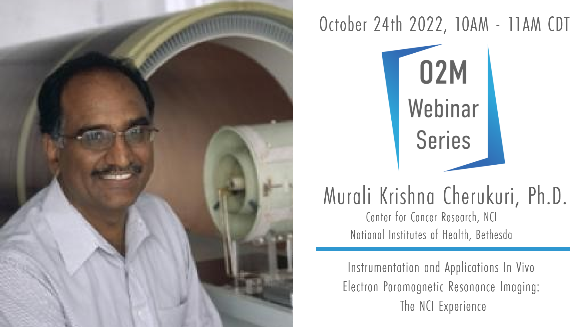
O2M Webinar: Instrumentation and Applications of In Vivo Electron Paramagnetic Resonance Imaging: The NCI Experience | Murali Krishna Cherukuri, Ph.D., NCI
Instrumentation and Applications In Vivo Electron Paramagnetic Resonance Imaging: The NCI Experience
Moderator: Dr. Periannan Kuppusamy, Ph.D., Dartmouth College
About the Speaker: Dr. Krishna, Murali, obtained a PhD from the Indian Institute of Technology in Madras, India, in 1984. He then joined the National Cancer Institute (NCI) as a Visiting Fellow in 1984. Since 1993, Murali has been a Principal Investigator, and Chief of the Biophysical Spectroscopy Section of the Radiation Biology Branch, Center for Cancer Research, NCI. In 2006, he was inducted into Senior Biomedical Research Service at NIH. His activities and accomplishments have directly related to the fields of radiation oncology and cancer biology. Over the years, Murali has published over 300 articles. Among the many honors and scientific recognitions awarded to Murali are the several Federal Technology Transfer Awards and an Invitational Visiting professorship with the Japan Society for Promotion of Science. He has been a member of the Editorial Board of Magnetic Resonance in Medicine since 2005 and is currently a member of the editorial board for the AACR journal Cancer Research. He is a member of the Interagency Group/National Security Agency for allocation of 3He. He was championing the allotment of 3He for medical imaging. He is the key member in the NCI clinical metabolic MRI. Murali is a major leader in EPR instrumentation development, testing, and biological applications of novel, non-invasive molecular/functional imaging modalities for potential clinical applications. His research has focused on two areas of molecular imaging: the measurement of tumor and normal tissue oxygen concentration, and hyperpolarized 13C-pyruvate metabolic imaging. Importantly, his pre-clinical research is expanding our knowledge of the tumor microenvironment with respect to tumor hypoxia and its clinical implications. He has a latitude of superb scientific judgment and balanced wisdom to make significant contributions in clinical and translation imaging in cancer. In 2014, was awarded the IES Silver Medal in Biology/Medicine in recognition of his outstanding contribution and dedication to advancing the biomedical application of EPR technology, translating basic research for clinical applications, and excellence in scientific research.
About the Webinar: The influence of molecular oxygen with its two unpaired electrons on a biocompatible paramagnetic spin probe’s spin-lattice (T1) and spin-spin (T2) relaxation processes allows the determination of pO2 in vivo non-invasively and quantitatively. EPR signals can be acquired through frequency-domain or time-domain modes of acquisition. EPR images can be generated by either projection-reconstruction using frequency encoding or phase-encoding of the spatial distribution. Obtaining the pO2 dependent line widths can be achieved by Spectral-spatial or T2-weighted approaches. An overview of the signal acquisition, image formation and reconstruction will be presented. Applications in pre-clinical models of human cancer will be presented.
